
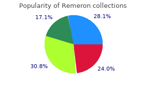
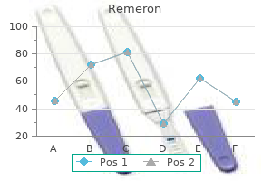
Remeron
"Remeron 15 mg purchase on line, treatment with cold medical term".
P. Kaffu, M.B. B.CH., M.B.B.Ch., Ph.D.
Co-Director, University of North Carolina School of Medicine
The sperm cells continue to mature inside the epididymis symptoms cervical cancer 30 mg remeron order visa, creating the capability to swim and the flexibility to bind to the oocyte medications ms treatment remeron 15 mg overnight delivery. Final modifications in sperm cells treatment lung cancer trusted remeron 30 mg, called capacitation (kapas i-ta shun), happen after ejaculation of semen into the vagina and previous to fertilization. The ductus deferens (duk tus def er-enz), or vas deferens, emerges from the epididymis and ascends alongside the posterior facet of the testis to turn into associated with the blood vessels and nerves that provide the testis. Each spermatic wire consists of the ductus deferens, testicular artery and veins, lymphatic vessels, and testicular nerve. Each ductus deferens extends, in the spermatic twine, via the stomach wall by method of the inguinal canal. Each ductus deferens then crosses the lateral wall of the pelvic cavity and loops behind the posterior floor of the urinary bladder to approach the prostate gland (figure 19. Blood vessels, including the dorsal artery and vein, and the dorsal nerve of the penis are seen. Corpus spongiosum Spongy urethra (b) Ventral floor Reproductive 536 Chapter 19 the ductus deferens is about 45 cm. Just earlier than reaching the prostate gland, the ductus deferens increases in diameter to turn into the ampulla of the ductus deferens (figure 19. The wall of the ductus deferens accommodates smooth muscle, which contracts in peristaltic waves to propel the sperm cells from the epididymis by way of the ductus deferens. Seminal Vesicle and Ejaculatory Duct Near the ampulla of every ductus deferens is a sac-shaped gland referred to as the seminal vesicle (semi-nal vesi-kl). A brief duct extends from the seminal vesicle to the ampulla of the ductus deferens. The ducts from the seminal vesicle and the ampulla of the ductus deferens be part of on the prostate gland to type the ejaculatory (e-jaku-la-tor-e) duct. Each ejaculatory duct extends into the prostate gland and ends by becoming a member of the urethra within the prostate gland (figure 19. The male urethra (u-re thra) extends from the urinary bladder to the distal finish of the penis (figure 19. The urethra can be divided into three elements: the prostatic urethra, which passes via the prostate gland; the membranous urethra, which passes via the ground of the pelvis and is surrounded by the external urinary sphincter; and the spongy urethra, which extends the size of the penis and opens at its finish. While male reproductive fluids are passing by way of the urethra, a sympathetic reflex causes the internal urinary sphincter to contract, which retains semen from passing into the urinary bladder and prevents urine from entering the urethra. Each is about 5 cm lengthy and tapers into a short duct that joins the ampulla of the ductus deferens to kind the ejaculatory duct, as previously mentioned. The prostate (pros tat) gland consists of both glandular and muscular tissue and is concerning the size and shape of a walnut (figure 19. His doctor reported reasonable enlargement of the prostate gland but detected no obvious tumorlike structures in a bodily examination. Urethra penis the penis (pe nis) is the male organ of copulation and functions within the switch of sperm cells from the male to the feminine. Engorgement of this erectile tissue with blood causes the penis to enlarge and become agency, a process known as erection (e-rek shun). Two columns of erectile tissue form the dorsal portion and the edges of the penis and are referred to as the corpora cavernosa (kor pora kav-er-nos a). The third, smaller erectile column occupies the ventral portion of the penis and is called the corpus spongiosum (kor pus spun-ge-o sum). It expands over the distal end of the penis to form a cap, the glans (glanz) penis. The spongy urethra passes through the corpus spongiosum, together with the glans penis, and opens to the exterior because the exterior urethral orifice. The pores and skin is firmly connected at the base of the glans penis, and a thinner layer of pores and skin tightly covers the glans penis. The skin of the penis, especially the glans penis, is nicely provided with sensory receptors. A free fold of pores and skin, referred to as the prepuce (pre pus), or foreskin, covers the glans penis (see figure 19. Glands Reproductive Predict 2 the seminal vesicles are glands consisting of many saclike constructions situated next to the ampulla of the ductus deferens (figure 19. Changes within the dimension and texture of the prostate gland could be a sign of developing prostate most cancers. Suggest a way that the dimensions and texture of the prostate gland can be examined by palpation with out surgical strategies (see figure 19. In younger adults, every is about the size of a pea, but they decrease in size with age. Hormones management the development of reproductive constructions, the development of secondary sexual characteristics, spermatogenesis, and some aspects of sexual habits. The mature neural mechanisms are primarily concerned in controlling the sexual act and within the expression of sexual habits. Semen (se men) is a mix of sperm cells and secretions from the male reproductive glands. The seminal vesicles produce about 60% of the fluid, the prostate gland contributes approximately 30%, the testes contribute 5%, and the bulbourethral glands contribute 5%. The bulbourethral glands and the mucous glands in the urethra produce a mucous secretion, which lubricates the urethra, helps neutralize the contents of the usually acidic urethra, provides a small quantity of lubrication throughout intercourse, and helps cut back acidity within the vagina. The thick, mucuslike secretion of the seminal vesicles accommodates the sugar fructose and different vitamins that nourish sperm cells. The seminal vesicle secretions also contain proteins that weakly coagulate after ejaculation and enzymes which may be thought to assist destroy abnormal sperm cells. Prostaglandins, which stimulate smooth muscle contractions, are present in high concentrations in the secretions of the seminal vesicles and might trigger contractions of the feminine reproductive tract, which assist transport sperm cells via the tract. The thin, milky secretions of the prostate have an alkaline pH and assist neutralize the acidic urethra, as well as the acidic secretions of the testes, the seminal vesicles, and the vagina. Prostatic secretions also comprise proteolytic enzymes that break down the coagulated proteins of the seminal vesicles and make the semen extra liquid. The normal quantity of semen is 2�5 milliliters (mL), with each milliliter of semen sometimes containing about one hundred million sperm cells. It additionally increases the secretion of a hormone referred to as inhibin (inhib in; to inhibit). In boys, puberty generally begins between the ages of 12 and 14 and is basically accomplished by age 18. Secondary sexual characteristics in males embody hair distribution and progress, skin texture, fat distribution, skeletal muscle development, and changes in the larynx. After puberty, testosterone maintains the adult construction of the male genitals, reproductive ducts, and secondary sexual characteristics. Testosterone enters sure cells inside the mind, particularly inside the hypothalamus, and influences their features. The blood ranges of testosterone stay relatively constant all through the lifetime of a male, from puberty until about 40 years of age. Thereafter, the degrees slowly decline to approximately 20% of this worth by eighty years of age, causing a sluggish lower in sex drive and fertility. Predict three effects of testosterone Testosterone (tes tos te-ron) is the major male hormone secreted by the testes. Testosterone influences reproductive organs and nonreproductive structures (table 19. During puberty, testosterone causes the enlargement and differentiation of the male genitals and the reproductive duct system. It is important for spermatogenesis and for the development of male secondary sexual characteristics. The secondary sexual characteristics are those structural and behavioral adjustments, apart from within the reproductive organs, that develop at puberty and distinguish males from females. Predict the impact on secondary sexual characteristics, exterior genitalia, and sexual behavior if the testes fail to produce regular amounts of testosterone at puberty. Reproductive the male sex act is a complex sequence of reflexes that result in erection of the penis, secretion of mucus into the urethra, emission, and ejaculation.
Additional information:
The authors discovered a survival profit in main restore of superior mesenteric venous injuries although they advocated rapid ligation in the unstable affected person afflicted by multiple other life-threatening injuries treatment ringworm remeron 30 mg order line. Expectedly medicine 853 order remeron 30 mg overnight delivery, ligation resulted in important bowel edema and venous engorgement with potential for splanchnic hypertension syndrome and bowel necrosis treatment efficacy remeron 15 mg generic mastercard. It is a renaismilitary extremity wounds: a preliminary outcome evaluation with 2-year sance that has favorably impacted patient morbidity and morfollow-up. J Vasc Surg 50(3):549�555, dialogue gies relating to vascular damage within the bigger context of trauma. As the applying and acceptemporary intravascular shunts at a civilian Level I trauma heart. Am J Surg 172(5): of move with short-term vascular shunts reduces circulating markers of 405�410, 1996. J Vasc Surg fifty two: ence gained in France during the Great War, 1914�1918, Bristol, 1919, 91�96, 2010. Eger M, Golcman L, Goldstein A: using a quick lived shunt in the ondary to native arterial harm after penetrating trauma. Hossny A: Blunt popliteal artery harm with complete decrease limb ischdrome in traumatic brachial artery injuries: an institutional experience emia: is routine use of temporary intraluminal arterial shunt justified Sriussadaporn S, Pak-art R: Temporary intravascular shunt in complicated tion shunting following penetrating trauma. Ding W, Ji W, Wu X, et al: Prolonged indwelling time of temporary vasarterial shunts without systemic anticoagulation. Am J Surg 180(6):493� cular shunts is associated with increased endothelial injury within the porcine 496, 2000. J Postgrad Med 38:68�69, ity throughout harm control: intraluminal shunting for proximal superior 1992. Johansen K, Bandyk D, Thiele B, et al: Temporary intraluminal shunts: Surg 181(5):519�522, 1975. With decreased transport occasions and knowledge of these past experiences, Rich and colleagues successfully applied arterial repair in the majority of patients in the Vietnam War and subsequently reported an amputation rate of 13%. In that experience, practically all interposition grafts have been reversed nice saphenous vein; and that type of reconstruction was utilized in 46% of the circumstances. Definition of Problem Identification of the Optimal Vascular Conduit the search for the optimum vascular conduit, in both elective and emergency conditions, has been a supply of debate and the supply of many analysis tasks. The perfect vascular conduit should be durable, resistant to infection, and readily accessible or available. In quite a few research of elective peripheral vascular bypass, autologous vein has confirmed superior to prosthetic modalities in the lower extremities, whereas prosthetic grafts are generally higher fitted to the bigger caliber central arteries. Unlike elective conditions, trauma instances differ in the sense that sufferers are usually youthful and have healthy vessels freed from atherosclerotic occlusive disease that can complicate repair. Specifically, the necessity to assure adequate softtissue protection to defend the conduit from contamination and disruption typically determines final success or failure. As documented all through this textual content, the method to vascular trauma is generally straightforward. Approaches to the injured vessel include major restore or restoration of perfusion utilizing an interposition or bypass graft. The strategy of patch angioplasty can be a helpful strategy in choose accidents which might be much less severe. Also, one should contemplate the diploma of ischemia more likely to outcome from vessel ligation. If the artery is minimally disrupted, it may be able to be d�brided, mobilized, and repaired primarily. When considering interposition or bypass grafting, one should handle the identical technical elements that are important in elective vascular reconstruction as follows: (1) influx vessel, (2) outflow vessel, and (3) conduit. Although the vascular harm itself could also be simple, the affected person is often not simple and will have suffered a number of injuries. The ease of availability and necessary size of conduit wanted are additionally elements to be thought-about when pursuing this type of reconstruction. It can be good to imagine that one answer applies to both army and civilian state of affairs, however the settings (and the nature of the wounds) are most frequently completely different. This chapter will describe the choices for selection of the vascular conduit to be used for restore of vascular damage. Types of Conduit using a vascular conduit in vascular trauma is, in principle, the same as its use for atherosclerotic occlusive or aneurysmal illness. The categories of vascular reconstruction are main repair, patch angioplasty, interposition (in situ) graft, and bypass graft. Extremity vascular injuries are mostly managed by using the interposition graft method with great saphenous vein as the most typical conduit. There is controversy over the position of prosthetic conduit within the setting of trauma, significantly in closely contaminated wounds with soft-tissue defects. Graft an infection resulting in thrombosis or anastomotic or graft disruption can happen with autologous vein or prosthetic conduits. Routing of grafts away from or out of the zone of contamination and assuring viable soft-tissue coverage diminishes this threat of graft-related issues. Research and improvement geared toward autogenous, tissue-engineered conduits could provide an "off-the-shelf" alternative to management of vascular trauma sooner or later. The degree of contamination can be minor similar to with a single stab wound or a laceration with a bit of glass, or it may be major similar to with an open femur fracture with softtissue wound. However, because of the complexities of different trauma situations similar to bilateral lower extremity damage, this conduit will not be possible or applicable. In uncommon cases, one could choose to use an arterial conduit for vascular reconstruction. Because the venous system has multiple redundant outflow tracts, there are several decisions for vein harvest. The lower extremity has the longest and mostly used options, together with the greater and lesser saphenous veins, the femoral vein, and even and dorsal foot vein. The cephalic and basilic veins of the higher extremity can be utilized independently or as a longer single phase graft. In the neck, the anterior, exterior, and inside jugular veins are options for vascular conduit. The veins of the neck are most commonly used as adjuncts for carotid artery repair because of their proximity. Use of autologous vein requires adhering to the tenants of secure and efficient dissection and procurement. In general, superficial veins may be harvested using a single steady incision, skip incisions, or a more moderen minimally invasive approach. The single incision is essentially the most expedient and mostly described approach for higher saphenous vein harvest. However, this is related to wound infection and dehiscence in 17% to 44% of patients. With this system the vein is harvested with electrocautery via a number of percutaneous incisions. Although threat of wound an infection is decreased with the endoscopic method, this does carry the added danger of thermal injury to the vein. Arterial conduits can also have improved dealing with traits, higher compliance match and even superior patency. The use of autologous arterial conduit is possible and efficacious but remains restricted in the setting of trauma as a outcome of the paucity of harvest sites, their challenging anatomic places, and the dearth of redundancy or length. The internal mammary (internal thoracic) artery is probably the most generally used arterial conduit. However, because of its confined location, entry is simply possible via a median sternotomy. The internal iliac artery can be utilized, but that is infrequent except in choose circumstances of pediatric injury. Klonaris et al described the benefits of using the inner iliac artery for restore of contaminated femoral artery pseudoaneurysm ensuing from trauma from repeated access throughout illicit drug use. This report describes using inside iliac artery for reconstruction in 9 (5 patch, four interposition graft) of 12 sufferers. At a imply of 19 months after restore, Klonaris et al reported no issues or cases of limb loss.
These shelves swing to a horizontal position and start to fuse with one another at about 56 days of growth medications zetia cheap remeron 15 mg free shipping. A cleft palate can range in severity from a slight cleft of the uvula to a fissure extending the complete size of the palate medicine emoji remeron 30 mg purchase with mastercard. Development of the organ techniques the main organ systems seem and begin to develop through the embryonic period (second to eighth week of development) symptoms after hysterectomy discount remeron 15 mg mastercard. This interval is therefore also known as the period of organogenesis (or gano-jen e-sis). While the neural tube is forming (18�26 days), the rest of the embryo is folding to type a tube alongside the higher part of the yolk sac (figure 20. The growing digestive tract pinches off from the yolk sac as a tube but stays attached in the middle to the yolk sac by a yolk stalk. General Features Fertilization, blastocyst Integumentary System Skeletal System Melanocytes type from neural crest. Nervous System Ectoderm endocrine System Ectoderm, mesoderm, endoderm Mesoderm thyroid begins to develop. Urinary System Reproductive System Mesoderm, endoderm Mesoderm, endoderm Reproductive Development, Heredity, and Aging 569 A considerable variety of outpocketings seem at about 28 days after fertilization along the complete length of the digestive tract (figure 20. A surprisingly giant variety of important internal organs develop from those outpocketings, together with the auditory tubes, tonsils, thymus, anterior pituitary gland, thyroid gland, parathyroid glands, lungs, liver, pancreas, and urinary bladder. The coronary heart develops from two blood vessels, which lie aspect by facet within the early embryo and fuse about 21 days after fertilization right into a single, midline coronary heart (figure 20. Blood vessels form from "blood islands" on the surface of the yolk sac and contained in the embryo. The single ventricle is subdivided into two chambers by the event of an interventricular (in-ter-ven-trik u-lar) septum (figure 20. An interatrial (in-ter-a tre-al) septum forms to separate the two atria (figure 20. An opening within the interatrial septum referred to as the foramen ovale (o-val e) connects the two atria and permits blood to flow from the proper to the left atrium within the fetus. Because of the foramen ovale, some blood within the fetus passes from the right atrium to the left atrium and bypasses the age (Days Since Fertilization) 31�35 Hand and foot plates on limbs sensory receptors seem in pores and skin. Mesoderm condensation in areas of future bone Muscle precursor cells enter limb buds. Cartilage in web site of future humerus 36�40 Fingers and toes appear; lips type; embryo 15 mm 41�45 External ear forming; embryo 20 mm Collagen fibers are clearly current in skin. Cartilage in website of future ulna and radius 46�50 Embryo 25 mm 51�55 Limbs elongate to adult proportions; embryo 35 mm Extensive sensory nerve endings in skin 56�60 Face is distinctly human in appearance. Cartilage in site of future hand and fingers Functional muscle ossification begins in clavicle and then in other bones. Tonsil Thymus and parathyroid glands Lung Pharynx Auditory tube Anterior pituitary Stomach Liver Embryonic kidney Pancreas Yolk stalk Intestine Urinary bladder Rectum the kidneys develop from mesoderm located alongside the lateral wall of the physique cavity (see determine 20. The embryonic kidneys are far more in depth than the grownup kidneys, extending the whole size of the physique cavity. They are carefully related to inner reproductive organs, such as the ovaries or testes, and reproductive ducts, such because the uterine tubes or ductus deferens. Most of the embryonic kidneys degenerate, with solely a really small part forming the adult kidney. Growth of the Fetus the embryo turns into a fetus about 8 weeks after fertilization (figure 20. The starting of the fetal period is marked by the beginning of bone ossification. In the embryo, a lot of the organ techniques are growing, whereas within the fetus the organs are current. The progress through the fetal interval represents more than a 15-fold increase in size and a 1400-fold improve in weight. Fine, gentle hair called lanugo (la-noo go) covers the fetus, and a waxy coat of free epithelial cells referred to as vernix caseosa (ver niks ka se-o sa) forms a protective layer between the fetus and the amniotic fluid. Subcutaneous adipose tissue that accumulates within the fetus provides a nutrient reserve, helps insulate, and aids the newborn in sucking by strengthening and supporting the cheeks, so that a small vacuum could be developed in the oral cavity. The foramen ovale usually closes off at the time of start, and blood then circulates by way of the best ventricle and the lungs. An interatrial septal defect or a ventricular septal defect normally leads to a heart murmur. Development, Heredity, and Aging 571 1 20 days after fertilization At this age, the guts consists of two parallel tubes that may fuse right into a single, midline coronary heart. Fusing coronary heart tube 1 Unfused coronary heart tubes Ventricle 2 2 22 days after fertilization the 2 parallel tubes have fused to type one tube. This tube bends because it elongates (blue arrows counsel the path of bending) throughout the confined area of the pericardium. Atrium Interatrial septum three Left atrium 3 31 days after fertilization the interatrial septum (green) and the interventricular septum grow towards the center of the heart. Right atrium Left ventricle Canals between atria and ventricles Right ventricle Interventricular septum four 4 35 days after fertilization the interventricular septum is almost complete. A foramen, which will turn out to be the left facet of the foramen ovale, opens in the left side of the interatrial septum (green) as the proper side of the interatrial septum begins to kind (blue). Interatrial septum Foramen Interventricular septum Interatrial septum 5 Final embryonic situation of the interatrial septum A foramen stays in the proper side of the interatrial septum (blue), which varieties the right part of the foramen ovale. Blood from the best atrium can move via the foramen ovale into the left atrium. After delivery, as blood begins to move in the other direction, the left facet of the interatrial septum is pressured towards the proper facet, closing the foramen ovale. Growth of the placenta essentially stops at about 35 weeks, limiting fetal growth. The average weight at this level is 3250 g (7 lb, 2 oz) for a female fetus and 3300 g (7 lb, four oz) for a male fetus. Near the top of pregnancy, the uterus turns into progressively extra excitable and usually displays occasional contractions that turn out to be stronger and more frequent till parturition is initiated. The cervix progressively dilates, and strong uterine contractions help expel the fetus from the uterus through the vagina. Labor is the period during which uterine contractions occur that result in expulsion of the fetus. Although labor may differ greatly from woman to woman and from one pregnancy to another for a similar lady, it can usually be divided into three stages. During this phase, the amnion surrounding the fetus ruptures, and amniotic fluid flows through the vagina to the exterior. This occasion is often referred to as the "water breaking" and often occurs naturally, but the amnion could must be ruptured artificially. The second stage of labor, usually called the expulsion phase, lasts from the time of maximum cervical dilation until the time the infant exits the vagina. The third stage of labor, usually referred to as the placental stage, entails the expulsion of the placenta from the uterus. Contractions of the uterus trigger the placenta to tear away from the wall of the uterus. Some bleeding from the uterine wall occurs due to the intimate contact between the placenta and the uterus. However, bleeding is normally restricted because uterine clean muscle contractions compress the blood vessels. Compare and contrast clinical age and developmental age for fertilization, implantation, the beginning of the fetal period, and parturition. However, estrogen ranges frequently increase within the maternal circulation, thrilling uterine clean muscle. Thus, the inhibitory influence of progesterone on clean muscle is overcome by the stimulatory effect of estrogen near the top of pregnancy. Oxytocin stimulates uterine contractions, which transfer the fetus farther into the cervix, inflicting further stretch. This positive-feedback mechanism stops after supply, when the cervix is not stretched.
Sometimes a person who has hyperventilated before swimming underneath water passes out whereas nonetheless beneath water and drowns medications requiring central line 15 mg remeron purchase with amex. Propose an explanation to account for the elevated pH values following the start of the race medications used to treat ptsd remeron 15 mg cheap amex. His mother took him to see his physician symptoms xanax treats purchase remeron 15 mg otc, who explained that Demondre should avoid dairy merchandise (which include the sugar lactose), especially cheese pizza! After studying the sections "Secretions of the Small intestine" and "Digestion, Absorption, and transport," clarify why Demondre can now not eat lactose without side effects. Enzymes within the digestive system break the particles down into very small molecules, that are absorbed into the circulation and transported everywhere in the physique. There, those molecules are damaged down by other enzymes to release energy or are assembled into new molecules to build tissues and organs. This chapter describes the construction and performance of the digestive organs and their accessory glands. During the method of digestion, food is damaged down from complicated particles to smaller molecules that could be absorbed. The epithelial cells that line the lumen of the small gut take in the small molecules of vitamins (amino acids, monosaccharides, fatty acids, vitamins, minerals, and water) that result from the digestive process. Undigested material, such as fiber from meals, plus waste merchandise excreted into the digestive tract are eradicated in the feces. It is a thick layer of unfastened connective tissue containing nerves, blood vessels, and small glands. In most parts of the digestive tract it consists of an internal layer of circular clean muscle and an outer layer of longitudinal smooth muscle. Another nerve plexus, additionally innervated by autonomic nerves, lies between the 2 muscle layers. Together, the nerve plexuses of the submucosa and muscularis compose the enteric (en-ter ik) nervous system. This nervous system, which is a division of the autonomic nervous system, is extraordinarily essential in controlling motion and secretion inside the tract (see chapter 8). The fourth, or outermost, layer of the digestive tract is both a serosa or an adventitia. The serosa consists of the peritoneum, which is a clean epithelial layer, and its underlying connective tissue. Regions of the digestive tract not coated by peritoneum are covered by a connective tissue layer called the adventitia (ad ven-tish a; overseas, coming from outside), which is steady with the encompassing connective tissue. Peritoneum the physique wall of the abdominal cavity and the abdominal organs is covered with serous membranes (figure sixteen. The serous membrane that covers the organs is the serosa, or visceral peritoneum (per i-to-ne um; to stretch over). The serous membrane that strains the wall of the abdominal cavity is the parietal peritoneum. Many of the organs of the abdominal cavity are held in place by connective tissue sheets referred to as mesenteries (mes en-ter-ez). Mesentery is a basic term referring to the serous membranes connected to the belly organs. The mesenteries consist of two layers of serous membranes with a skinny layer of unfastened connective tissue between them. The mesentery connecting the lesser curvature of the stomach to the liver and diaphragm known as the lesser omentum (o-men tum), and the mesentery connecting the larger curvature of the stomach to the transverse colon and posterior body wall is recognized as the larger omentum. Adipose tissue accumulates in the greater omentum, giving it the looks of a fat-filled apron that covers the anterior surface of the abdominal viscera. The mesentery that attaches the small gut to the posterior belly wall is called the mesentery proper. Because the digestive tract is open on the mouth and anus, the inside of the tract is continuous with the skin surroundings, and food coming into the digestive tract might comprise not solely useful vitamins but also indigestible components such as fiber, or harmful supplies similar to bacteria. Therefore, the internal lining of the digestive tract serves as a protecting barrier to those indigestible and harmful supplies and vitamins have to be particularly transported across the wall of the digestive tract. Once throughout the wall of the digestive tract, the vitamins enter the circulation to access tissues of the physique. The digestive tract consists of the oral cavity, pharynx, esophagus, abdomen, small intestine, large intestine, and anus. The salivary glands empty into the oral cavity, and the liver and pancreas are related to the small gut. These are the mucosa, the submucosa, the muscularis, and a serosa or an adventitia (figure 16. The innermost tunic, the mucosa (mu-ko sa), consists of mucous epithelium, a free connective tissue called the lamina propria, and a skinny clean muscle layer, the muscularis mucosae. The epithelium in the mouth, esophagus, and anus resists abrasion, and the epithelium within the stomach and gut absorbs and secretes. Digestive If you placed a pin completely through each folds of the larger omentum, via what number of layers of easy squamous epithelium would the pin move The retroperitoneal organs embody the duodenum, pancreas, ascending colon, descending colon, rectum, kidneys, adrenal glands, and urinary bladder. The lips are muscular structures, fashioned mostly by the orbicularis oris (or-biku-laris oris) muscle (see figure 7. The keratinized stratified epithelium of the pores and skin becomes skinny on the margin of the lips. The color from the underlying blood vessels can be seen through the skinny, transparent epithelium, giving the lips a reddish-pink appearance. At the inner margin of the lips, the epithelium is steady with the moist stratified squamous epithelium of the mucosa in the oral cavity. They assist manipulate the food throughout the oral cavity and maintain the food in place while the enamel crush or tear it. Mastication begins the process of mechanical digestion, which breaks down large food particles into smaller ones. The anterior a part of the tongue is relatively free, except for an anterior attachment to the ground of the mouth by a skinny fold of tissue called the frenulum (frenu-lum) (figure 16. The anterior two-thirds of the tongue is covered by papillae, a few of which include style buds (see chapter 9). The posterior one-third of the tongue is devoid of papillae and has only some scattered taste buds. In addition, the posterior portion does include a appreciable quantity of lymphatic tissue, which helps kind the lingual tonsil (see chapter 14). The tongue moves meals within the mouth and, in cooperation with the lips and cheeks, holds the meals in place during mastication. In addition, the tongue is a serious sensory organ for taste, in addition to one of the major organs of speech. Teeth There are 32 tooth in the normal grownup mouth, positioned within the mandible and maxillae. The teeth can be divided into quadrants: right higher, left upper, right decrease, and left decrease. In adults, every quadrant contains one central and one lateral incisor (in-si zor; to cut); one canine (ka nin; dog); first and second premolars (premo larz; molaris, a millstone); and first, second, and third molars (mo larz). The third molars are referred to as knowledge teeth as a result of they often appear in the late teens or early twenties, when the person is sufficiently old to have acquired a point of knowledge. Most of them are replacements for the 20 primary tooth, or deciduous (de-sid u-us) enamel, additionally known as milk or baby tooth, that are lost during childhood (figure sixteen. Each tooth consists of a crown with one or more cusps (points), a neck, and a root (figure 16. The center of the tooth is a pulp cavity, which is filled with blood vessels, nerves, and connective tissue, referred to as pulp. The pulp cavity is surrounded by a dwelling, mobile, bonelike tissue referred to as dentin (den tin; dens, tooth). The dentin of the tooth crown is covered by a particularly onerous, acellular substance referred to as enamel, which protects the tooth in opposition to abrasion and acids produced by micro organism in the mouth. The floor of the dentin in the root is roofed with cementum (semen tum), which helps anchor the tooth within the jaw. The enamel are rooted inside alveoli (al-ve o-li; sockets) alongside the alveolar processes of the mandible and maxillae.